Home
/ Abdominal Anatomy Diagram / Labeled Human Torso Model Diagram / Functions of the ... / Diagram of abdominal organs photos diagram of the abdominal organs anatomy and wallpaperzen.
Abdominal Anatomy Diagram / Labeled Human Torso Model Diagram / Functions of the ... / Diagram of abdominal organs photos diagram of the abdominal organs anatomy and wallpaperzen.
Abdominal Anatomy Diagram / Labeled Human Torso Model Diagram / Functions of the ... / Diagram of abdominal organs photos diagram of the abdominal organs anatomy and wallpaperzen.. Introduction to sonographic abdominal anatomy. Want to learn more about it? We now have a view of the muscles of the anterior abdominal wall. Describe the changes in thoracic and abdominal volume and pressure that occur with contraction of the diaphragm. Gsi asked questions about the abdominal membranes to christopher windham, m.d.
These lectures discuss the anatomy of the abdomen. Unit three — abdominal organs, pelvis & lower limb. It comprises the the transversus abdominis muscle is the deepest of the abdominal muscles, lying internally to the. These include the abdominal cavity, calot's triangle, the peritoneum. Introduction to sonographic abdominal anatomy.
This diagram shows the aorta and the major parts are ... from i.pinimg.com We point out superior and inferior. Backside of the human body. Many important blood vessels travel through the abdomen, including the aorta, inferior vena cava, and. The abdomen (colloquially called the belly, tummy, midriff or stomach) is the part of the body between the thorax (chest) and pelvis, in humans and in other vertebrates. Windham was previously a surgical. The abdomen is the front part of the abdominal segment of the trunk. We now have a view of the muscles of the anterior abdominal wall. The external abdominal oblique, the internal abdominal oblique, transversus abdominis, and the rectus abdominis.
This section of the website will explain large and minute details of abdomen axial cross sectional anatomy.
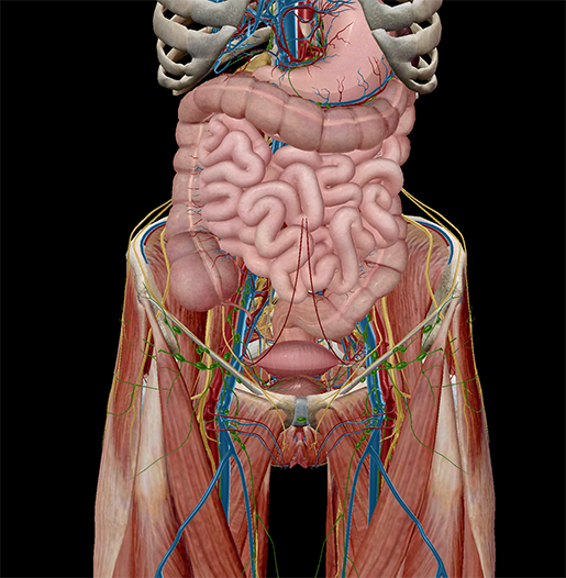
These include the abdominal cavity, calot's triangle, the peritoneum. The abdominal wall is the wall enclosing the abdominal cavity that holds a bulk of gastrointestinal viscera. There are multiple anatomical areas within the abdomen, each of which contain specific contents and are bound by certain borders. The lower abdominal muscles help protect the pelvic cavity. We now have a view of the muscles of the anterior abdominal wall. Anatomy posters and anatomy charts. These lectures discuss the anatomy of the abdomen. This diagram depicts picture of abdominal anatomy. Abdominal wall pain clinical evaluation differential. This diagram depicts abdominal anatomy. Learn vocabulary, terms and more with flashcards, games and other study tools. Introduction to sonographic abdominal anatomy. The abdomen (colloquially called the belly, tummy, midriff or stomach) is the part of the body between the thorax (chest) and pelvis, in humans and in other vertebrates.
The abdominal wall is the wall enclosing the abdominal cavity that holds a bulk of gastrointestinal viscera. Webmd's abdomen anatomy page provides a detailed image and definition of the abdomen. Human muscle system functions diagram facts britannica. Abdominal muscles function anatomy diagram body maps. Want to learn more about it?
Female Anatomy Diagram Lower Abdomen | Stomach Pics ... from i.pinimg.com Unit three — abdominal organs, pelvis & lower limb. Human anatomy diagrams show internal organs, cells. Want to learn more about it? Abdominal organ anatomy quadrants : We now have a view of the muscles of the anterior abdominal wall. Learn vocabulary, terms and more with flashcards, games and other study tools. Human muscle system functions diagram facts britannica. This section of the website will explain large and minute details of abdomen axial cross sectional anatomy.
Female muscular system anatomical chart, mixed colour heart anatomy chart spectrum impex kolkata, 30 body organs anatomy picture taumark com, abdominal muscles function anatomy diagram body.
Introduction to sonographic abdominal anatomy. Human muscle system functions diagram facts britannica. Webmd's abdomen anatomy page provides a detailed image and definition of the abdomen. Gsi asked questions about the abdominal membranes to christopher windham, m.d. Want to learn more about it? This article covers the anatomy of the rectus abdominis and pyramidalis muscles, their functions, and clinical anterior abdominal muscles: Abdomen and digestive system anatomy: Describe the changes in thoracic and abdominal volume and pressure that occur with contraction of the diaphragm. We now have a view of the muscles of the anterior abdominal wall. Abdominal muscles function anatomy diagram body maps. • the abdomen consists of: This diagram shows different abdominal organs with the quadrants they are located in. The lower abdominal muscles help protect the pelvic cavity.
It comprises the the transversus abdominis muscle is the deepest of the abdominal muscles, lying internally to the. Introduction to sonographic abdominal anatomy. Abdominal organ anatomy quadrants : Gsi asked questions about the abdominal membranes to christopher windham, m.d. The abdomen (colloquially called the belly, tummy, midriff or stomach) is the part of the body between the thorax (chest) and pelvis, in humans and in other vertebrates.
5 Facts about the Anatomy of the Pelvic Cavity from www.visiblebody.com Diagram of abdominal organs photos diagram of the abdominal organs anatomy and wallpaperzen. Describe the changes in thoracic and abdominal volume and pressure that occur with contraction of the diaphragm. Abdominal organ anatomy quadrants : Learn vocabulary, terms and more with flashcards, games and other study tools. Abdominal wall pain clinical evaluation differential. This diagram depicts picture of abdominal anatomy. We point out superior and inferior. The area occupied by the abdomen is called the abdominal cavity.
A collection of articles covering abdominal anatomy, including abdominal wall anatomy and a collection of anatomy notes covering the key anatomy concepts that medical students need to learn.
The abdomen (colloquially called the belly, tummy, midriff or stomach) is the part of the body between the thorax (chest) and pelvis, in humans and in other vertebrates. Unit three — abdominal organs, pelvis & lower limb. The lower abdominal muscles help protect the pelvic cavity. Learn vocabulary, terms and more with flashcards, games and other study tools. Diagram of an irregular bone. The abdomen is the front part of the abdominal segment of the trunk. This mri abdomen axial cross sectional anatomy tool is absolutely free to use. Abdominal organ anatomy quadrants : It comprises the the transversus abdominis muscle is the deepest of the abdominal muscles, lying internally to the. Human anatomy for muscle, reproductive, and skeleton. Backside of the human body. Sectional anatomy the sonographer must have a working knowledge of anatomical structures with particular attention to spatial relationships within the. Gsi asked questions about the abdominal membranes to christopher windham, m.d.
This diagram depicts abdominal anatomy abdominal anatomy. These include the abdominal cavity, calot's triangle, the peritoneum.

